Physiotherapy · Knee
Anterior Cruciate Ligament Tear: Complete Clinical Guide
An ACL injury is one of the most feared in sport and at work. Understanding its anatomy, grades of involvement and the fundamental role of physiotherapy is key to making the best therapeutic decisions.
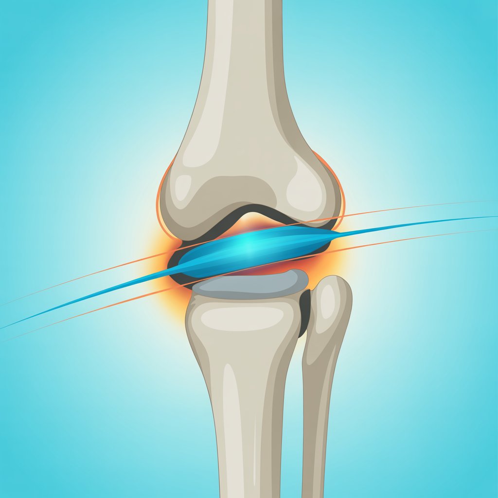
The knee is the largest joint in the human body and one of the most vulnerable to trauma and overload. The ligaments that stabilise it — the anterior cruciate ligament (ACL), posterior cruciate ligament (PCL), medial collateral ligament (MCL) and lateral collateral ligament (LCL) — work together to guarantee its function. When one of them is damaged, the mechanical balance of the entire joint is compromised.
The anterior cruciate ligament is the most frequently injured stabiliser. It sits inside the joint itself, connecting the posterior part of the femur to the anterior part of the tibia. Its main role is to prevent the tibia from sliding forward on the femur and to control rotational movements of the knee.
Anatomy and function of the ACL
Understanding the ACL's function is essential for understanding why its injury is so disabling. This ligament acts as the primary restraint to anterior tibial displacement and as a secondary brake to internal rotation. When it ruptures, instability occurs that causes progressive wear of the meniscus and articular cartilage.
Higher incidence in women
ACL tears are more common in women than men, due to biomechanical, hormonal and intercondylar notch angle differences.
Highest-risk sports
Football, basketball, skiing, volleyball and any sport involving sudden changes of direction, abrupt stops or jump landings.
Associated injuries
An ACL tear frequently accompanies injury to the medial meniscus or medial collateral ligament — the so-called "unhappy triad".
Injury mechanisms and symptoms
Most ACL tears occur without direct contact. The most common mechanisms involve knee hyperextension, a sudden pivot with the foot planted on the ground, or an abrupt stop during a run.
- Sensation of an audible "crack" or "pop" at the moment of injury
- Intense pain and rapid swelling of the knee in the first hours
- Difficulty walking due to a feeling of instability or the knee "giving way"
- Loss of range of motion in both flexion and extension
- Haemarthrosis (blood accumulation in the joint) from ligament bleeding
With a knee that swells rapidly after trauma, go to A&E to rule out a complete tear and assess whether other structures are affected using X-ray and, if appropriate, MRI.
First aid: the RICE protocol
In the moments immediately after the injury, correct action can reduce secondary damage and better prepare the joint for subsequent treatment:
Surgery or conservative treatment?
One of the most important decisions after an ACL tear is whether to proceed with surgery or opt for conservative treatment. This decision depends on multiple factors and must always be individualised.
«The therapeutic decision must consider the severity of the injury, the presence of associated structural damage, the patient's activity level, age and degree of functional instability.»
— Based on musculoskeletal physiotherapy clinical protocolsSurgical reconstruction options
Well-established technique. Uses the central third of the patellar tendon with bone blocks at both ends. Excellent long-term results.
Uses the gracilis and semitendinosus tendons. Less postoperative pain, although the ligamentisation process may be slightly slower.
No tissue is taken from the patient, reducing postoperative pain. Slightly higher infection risk and longer integration time.
The role of physiotherapy in recovery
Whether surgery or conservative treatment is chosen, physiotherapy is the fundamental pillar of recovery. The goal is not only that the knee stops hurting: it is to restore strength, stability, neuromuscular control and the patient's confidence in their joint.
- 1Initial phase: pain and swelling control
Cryotherapy, analgesic electrotherapy, gentle manual therapy and progressive joint mobility exercises. The goal is to restore range of motion and reduce early muscle atrophy.
- 2Strengthening phase
Closed kinetic chain exercises (leg press, assisted squat, step) to protect the graft and build quadriceps and hamstrings. Stationary cycling and swimming are progressively introduced.
- 3Neuromuscular re-education and proprioception
Balance, coordination and joint stability training using unstable surfaces, single-leg exercises and full lower limb kinetic chain work.
- 4Functional and sport return
Exercises specific to the patient's sport or activity: running, direction changes, jumps and stops. Strength symmetry and psychological confidence are assessed before authorising full return.
Prognosis and recovery times
The prognosis for an ACL tear is favourable when addressed correctly. After surgical reconstruction, average rehabilitation lasts between 7 and 9 months, although patients can continue improving for up to a year after the procedure.
It is essential not to rush the return to activity: a relapse can compromise additional structures and significantly complicate the prognosis. Sport return must be based on objective criteria of strength and stability, not just time elapsed.
Suffered a knee injury?
We assess you at Fisioin20, our clinic in the Salamanca district (C/ Don Ramón de la Cruz, 36, Madrid), with or without a prior diagnosis.
Request assessment →